Spotting a deadly melanoma often takes a handheld device. Does your doctor have one?
Published in News & Features
Lather on the sunscreen this holiday weekend and pay close attention during your skin exam.
That brown spot on your leg could look like nothing to worry about, particularly to the naked eye. But early skin cancer, including aggressive melanoma, could be missed with just a visual exam.
A recent viral headline illustrates the risk: “Mom, 37, Dies of Skin Cancer After Doctors Said Her Mole Was ‘Nothing to Worry About.’”
Josie Thompson’s melanoma, originating from a mole on her back, was initially dismissed by doctors despite her concerns and the mole regrowing multiple times. Thompson, from Plymouth, England, had formed a Facebook group to encourage others to get skin checks.
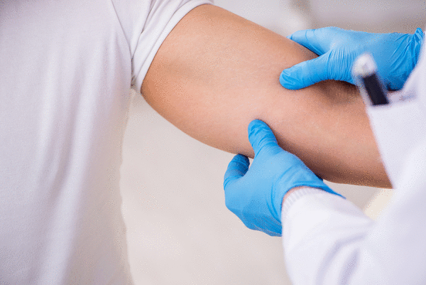
A new campaign is calling on more doctors, particularly in states like Florida with high rates of skin cancer, to use dermatoscopes for all skin exams. They suggest that patients seek dermatologists who use a dermatoscope, a small, handheld device that visualizes structures beneath the skin’s surface. Experts liken it to using a mammogram to detect breast cancer. The device is not new, but it has only recently been the focus of more training by medical schools and medical associations.
“It is something that we as dermatologists should use for every skin check,” said Dr. Natalia Jaimes, an associate professor at the University of Miami Medical School’s Dr. Phillip Frost Department of Dermatology & Cutaneous Surgery. “It increases our diagnostic accuracy to detect early melanomas.”
Melanoma is the most invasive skin cancer with the highest risk of death. It can even form under your fingernail.
While many primary care doctors, nurse practitioners and physician assistants do skin exams, few are trained in how to use a dermatoscope. The same is true of the older generation of dermatologists and some new dermatology residents.
Dr. Michael Christopher, an Arizona dermatologist, says his consistent dermoscopy use leads to skin cancer detection rates far above the national average. He says his goal, through public outreach, is to make dermoscopy a baseline tool, like a stethoscope.
He makes his case on his Instagram: “This spot is under 2 mm, on the leg, and looks normal. To the naked eye, it blends in. Only dermoscopy shows the features that make the diagnosis clear. If dermoscopy isn’t used routinely during a skin exam, lesions like this are easy to overlook. Awareness changes outcomes. Early detection changes lives.”
During an exam, Jaimes, also a dermatologist at Miami’s Sylvester Comprehensive Cancer Center, noticed a brown dot behind her patient’s ear that looked like a freckle. Peering into her dermatoscope, she saw beneath the skin’s surface, revealing patterns that characterize melanoma. She said it takes more than just using the device to detect skin cancer. “It requires training.”
In Florida, the need for skin cancer detection is great: Roughly 9,000 people a year in Florida get diagnosed with melanoma, and about 700 people annually in the state die from it, according to Florida Department of Health data. Melanoma is the most dangerous form of skin cancer because it is more likely to spread to other parts of the body.
A 2024 study published in JAMA Dermatology shows the difference a dermatoscope can make: Experienced dermatologists (those with over two years of practice) were nearly six times better at identifying melanoma when using dermoscopy compared with visual examination alone. Even more striking, dermatologists were more than 13 times as likely as primary care physicians to accurately diagnose melanoma using dermoscopic imaging.
Melanoma expert Dr. Maria Wei, a contributor to the study, said some of those skin cancers may eventually have been identified, but just a little later.
Wei, a professor of dermatology at the University of California, San Francisco, said she took a nationwide training course later in her career to learn how to use a dermatoscope. “I think that with time, the entire U.S. dermatology residency programs will be training in demoscopy,” she said.
The dermatoscope, which magnifies lesions, serves another purpose, too, said Dr. Andrew Miner, a Brevard County dermatologist, Mohs surgeon, and past president of the Florida Academy of Dermatology. “It prevents biopsies that don’t need to be done.”
Miner said he just treated a patient with skin cancer on the face. “I took care of that and he said, ‘I’ve got this spot on my wrist too.’ And it was kind of difficult to assess with the naked eye, and I got my dermatoscope out, and I looked at it, and I saw that it was a benign growth … so I got to leave it alone.”
The vast majority of lesions dermatologists examine with the dermatoscope are benign, Wei notes. “Fewer needless biopsies means we can also eliminate the uncertainty and anxiety that goes hand-in-hand with waiting to hear if you’ve got skin cancer.”
Wei said if you’re someone with several melanoma risk factors, like red hair, a family history of the disease, a high mole count, or blistering sunburns in your past, you would likely benefit from frequent monitoring with a dermatoscope.
If caught early, the melanoma survival rate has improved with breakthrough advancements in immunotherapy and targeted therapies.
“The trajectory is much different from years ago,” Jaimes said. She said anyone living in sunny Florida should check their skin regularly.
Anything new that lasts more than three or four weeks or anything changing — regardless of whether it is pink brown or black — should be brought to a doctor’s attention, she said. “As dermatologists, we need to do our part,” she said. “But everyone should advocate for themselves.”
_____
©2026 South Florida Sun-Sentinel. Visit sun-sentinel.com. Distributed by Tribune Content Agency, LLC.


Comments