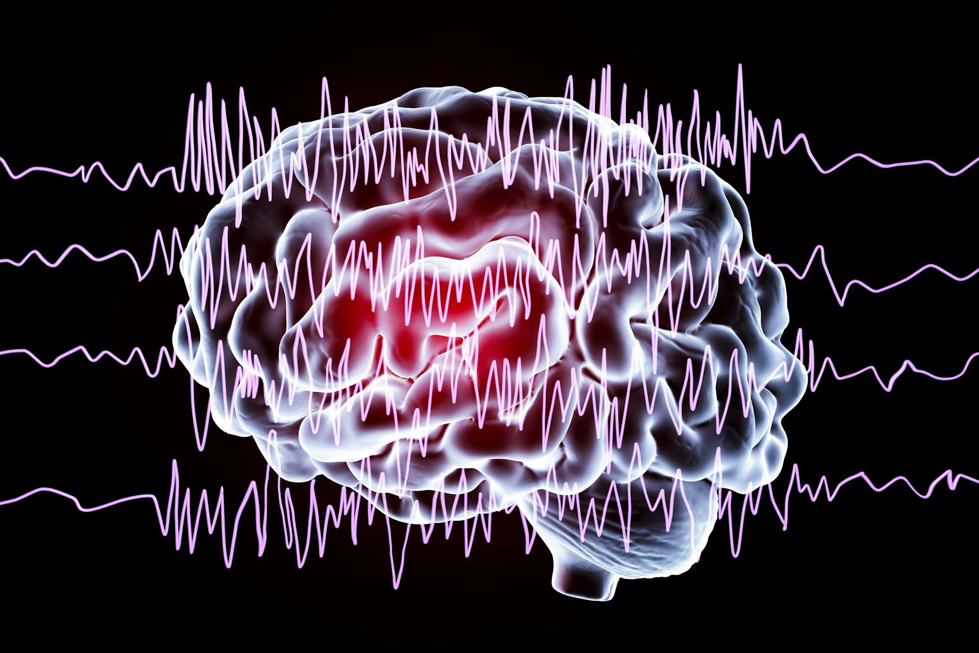
A century after the EEG was discovered, it remains a crucial tool for understanding the brain
Published in Health & Fitness

Jena, Germany, 1924: Working in near-isolation and with painstaking tediousness, the psychiatrist Hans Berger observes rhythmic electrical activity from the scalp of human subjects. He is convinced the activity arises from within the brain and coins the term “electroencephalogram.”
It is 10 years before the scientific community accepts Berger’s work, birthing the field of electroencephalography, or EEG for short.
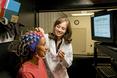
Today, the electroencephalogram – also abbreviated as EEG – is widely known as a medical test measuring brain electrical activity that’s used on patients who have, or are suspected to have, neurological disorders. The EEG provides a window into the living brain, with a continuous electrical readout of what is happening inside our heads. The procedure may be short, often just a 30-minute recording. But for patients monitored for diagnosis or treatment of brain disease, it can be continued for much longer – days or even weeks.
As a neurologist specializing in epilepsy, I use EEG on a daily basis. Our team at the University of Florida interprets thousands of EEGs a year in neurological patients. I also run a research lab where our goal is to understand the basic structure of the EEG in health and disease.
The story of EEG is colorful and littered with fables. Berger’s interest in brain electricity was not to combat disease, though that was his day job as a physician, but to find a biological basis for his belief in telepathy. He wondered whether the brain waves of EEG could convey thoughts through space, allowing people to read each other’s minds. He was unsuccessful in his mission, but the field he founded nonetheless took off.
By the mid-1930s, researchers had observed the striking differences between the awake and asleep EEG. The EEGs of patients with brain disease turned up a variety of unprecedented observations.
And then came a defining moment for modern medicine. In December 1934, a group of Boston physicians observed the rhythmic EEG spike-wave appearance of seizures in patients with “petit mal” epilepsy. Petit mal is an anachronistic term for a type of epilepsy where a patient’s flow of thought, speech or action momentarily freezes during seizures. For the first time, the symptoms and behavior of patients during seizures were correlated to a brain signal occurring in lockstep.
EEG quickly evolved from a scientific curiosity to a mainstream clinical tool. The first clinical EEG laboratory was set up at Massachusetts General Hospital in 1937. The practice grew in the ensuing decades into the specialized services that institutions like ours have offered since the 1970s.
So what, exactly, is the EEG?
Imagine taking two small metallic discs connected by a conducting wire. Place one disc on the scalp and connect the other to a neutral reference, such as the ear. Watch a tiny alternating current flow in the wire, proportional to the electrical activity sensed by the conducting contact. This activity is the EEG, the electrical milieu that bathes brain tissue.
...continued


Comments